The Spiralist
Why do helical seashells resemble spiralling galaxies and the human heart? Kevin Dann leads us into the gyre of James Bell Pettigrew’s Design in Nature (1908), a provocative and forgotten exploration of the world’s archetypal whorl.
August 5, 2021
 Scroll through the whole page to download all images before printing.
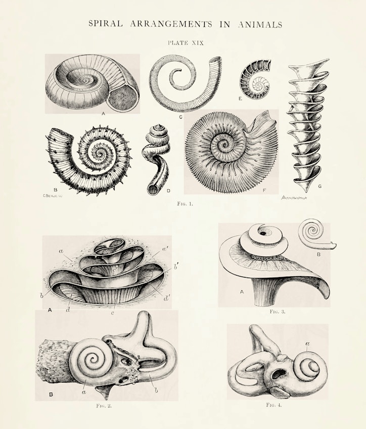
Plate XIX from the first volume of Pettigrew’s Design in Nature (1908), illustrating the resemblance between spiral shell formations and bony portions of the inner ear — Source.
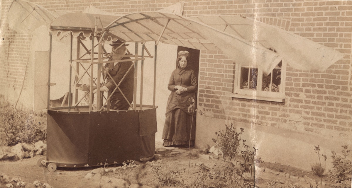
One halcyon spring day in 1903, the sixty-nine-year-old anatomist and naturalist Dr. James Bell Pettigrew sat at the top of a sloping street on the outskirts of St. Andrews, Scotland, perched inside a petrol-powered aëroplane of his own design. Over the course of forty years, ever since he began his aeronautical experiments in London in 1864, Pettigrew had constructed dozens of working models of various flying apparatus. From anatomical dissection and observations of animals in the wild and at the London Zoo, Pettigrew had come to conceive of all creatures — whether on land, in water, or in the air — as propelling themselves by throwing their bodies into spiraling curves, such that their movements were akin to waves in fluid, or to waves of sound. Instead of driving the wings vertically as in other flying machines modeled on animal flight, Pettigrew’s “ornithopter” emulated the movement that he had discovered to be universal in flying creatures: rhythmic figure-of-eight curves. To permit this undulatory motion, Pettigrew had furnished the root of the wing with a ball-and-socket joint; to regulate the several movements of the vibratory wing — comprised of bamboo cane from which issued tapering rods of whalebone covered in a thin sheet of India rubber — he employed a cross-system of elastic bands. A two-stroke engine’s piston drove this elaborate apparatus of helical biological mimicry.
 Scroll through the whole page to download all images before printing.
Pettigrew and a version of his ornithopter; the woman may be Elsie Bell Pettigrew (née Gray), philanthropist, founder of the Bell Pettigrew museum of natural sciences, and his wife — Source © The Royal Aeronautical Society (National Aerospace Library) / Mary Evans Picture Library.
The ornithopter covered a distance of about twenty meters during its maiden flight before crashing, breaking both the contraption’s spiral whalebone wings and its pilot’s own spiral hip. Convalescence gave Dr. Pettigrew the opportunity to begin work on Design in Nature: Illustrated by Spiral and Other Arrangements in the Inorganic and Organic Kingdoms as Exemplified in Matter, Force, Life, Growth, Rhythms, &c., Especially in Crystals, Plants, and Animals. In January 1908, as he was nearing its completion, Pettigrew looped back at the work’s end to reiterate what he had stated so vociferously at the beginning — the absolute primacy of design by a “Great First Cause” and “Omni–Present Framer and Upholder of the Universe”. After a lengthy essay considering the antiquity of man — and once again stressing that the human physical form had altered not at all for at least some ten thousand years — he concluded:
Man is not in any sense the product of evolution. He is not compounded of an endless number of lower animal forms which merge into each other by inseparable gradations and modifications from the monera up to man. . .
He is the highest of all living forms. The world was made for him and he for it. . . Everything was made to fit and dovetail into every other thing. . . There was moreover no accident or chance. On the contrary, there was forethought, prescience, and design.1
 Scroll through the whole page to download all images before printing.
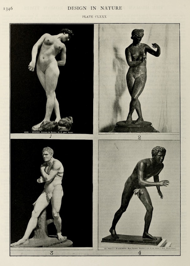
Plate CLXXX from the third volume of Pettigrew’s Design in Nature (1908), illustrating, via classical and modern sculpture, “diagonal screwing movements” that occur during “walking, swimming, and flying”. In numerical order: a life study by Gérome; Venus of Ostia; the Greek boxer Damoxenus by Canova; and Discobolus in bronze — Source.
Across the 3 volumes, 1416 pages, and nearly 2000 illustrations that made up his magnum opus Design in Nature, Dr James Bell Pettigrew barely mentioned Charles Darwin’s theory of natural selection, which he found “lame, halting, and impotent”.2 Though Pettigrew deeply admired the English naturalist — who had on more than one occasion (as had T. H. Huxley, Richard Owen, John Lubbock, St. George Mivart, and dozens of other leading men of science) visited Pettigrew at London’s Hunterian Museum of the Royal College of Surgeons of England to view his state-of-the-art anatomical and physiological preparations — Darwin seemed to Pettigrew only tentatively confident of the very theory he had proposed to explain Nature’s “endless forms most beautiful”.3 He expected that, within a generation, few would recall Darwinism as anything other than a passing fancy. What Pettigrew could not excuse were the egregious errors in Darwin’s pronouncement about the spiraling motions of Clematis, Convolvulus, Honeysuckle, Hops, and many other plants. Whatever positive contributions the retiring naturalist had made with his research on twining plants were undermined by his inexact language and thinking. Pettigrew strenuously objected to Darwin’s use of the term “reflex action” for these plants’ behavior, since this was a phrase used for action in nervous systems — of which Clematis, Convolvulus, and their cousins possessed none.4
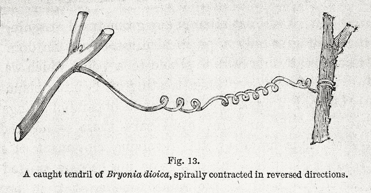
By way of a few ingenious experiments — conducted back in 1865, after reading Darwin’s “On the Movements and Habits of Climbing Plants” — Pettigrew had utterly demolished its author’s “irritability theory” for the movement of the green chimeras. Just as with spiral teeth, claws, horns, muscles, and bones, spirally-turning plant tendrils were in no way the result of external contact. These whirling, twirling structures, as free of contact as the ocean-suspended spiraling egg cases of sharks and dogfish, danced to some wholly invisible music.
 Scroll through the whole page to download all images before printing.
Illustration from Darwin’s “On the Movements and Habits of Climbing Plants”, accompanying the “irritability theory” that Pettigrew denounced — Source.
 Scroll through the whole page to download all images before printing.
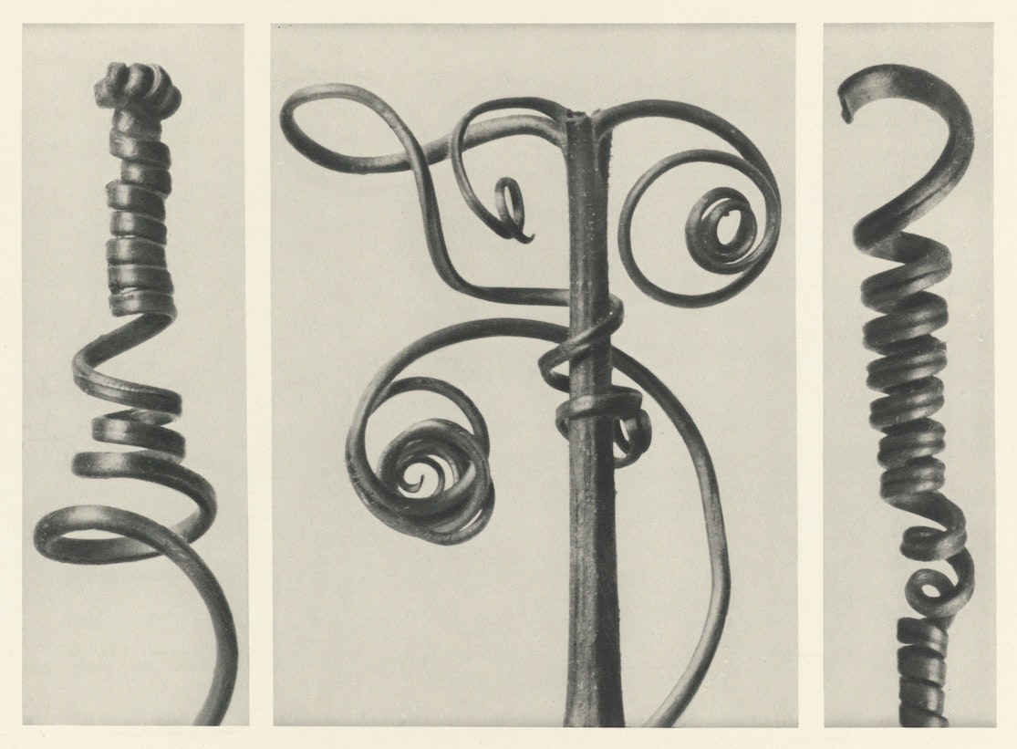
Tendrils of pumpkin (Cucurbita) under magnification. From Karl Blossfeldt’s 1928 Urformen der Kunst (Art Forms in Nature) — Source.
Pettigrew confessed himself totally spellbound by the mystery of Nature’s most ubiquitous, liquid, and quixotic form — the spiral. Though he had scrutinized this universal cipher from the macrocosmic spiral nebulae down to the dextro- and sinistro-helical microcosmic molecules of the periodic table, Pettigrew was left baffled by the question of its origin. The best that he could say was that the answer “by no means lies on the surface”.5 Overwhelmed as he was by the world’s archetypal whorl, he was sure of one thing — these marvelous spiral arrangements could not be of purely physical origin. A reviewer of Pettigrew’s Lancet series on circulation in plants and animals declared that the distinguished anatomist was a “spiralist” who found that organs were not only constituted spirally, but that they functioned spirally too.6
 Scroll through the whole page to download all images before printing.
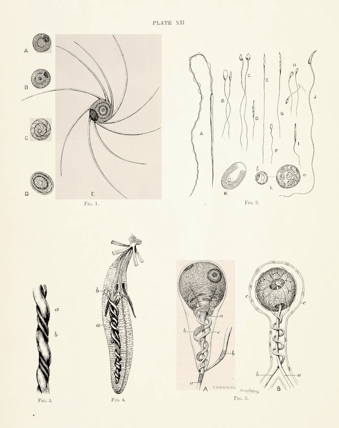
Plate XII from the first volume of Pettigrew’s Design in Nature (1908), illustrating “spiral formations and structures in spermatozoids, umbilical cord, intestine, and nerve cells” — Source.
Putting Isaac Roberts’ astonishing 1888 photographs of the Great Nebula in Andromeda and other spiral nebulae to cosmic effect at his argument’s outset, Pettigrew then immersed the reader in a cascade of more humble spiral forms — the mineral prochlorite; ram’s horns; bacteria from the River Thames; fossil algae carpogonia; dozens of figures of spiral fronds, floral bracts, stems, leaves, tendrils, and seeds in plants. Design in Nature’s plates of spirals in the animal world started with spermatozoa (of crayfish, rabbit, field mice, wood shrike, goldfinch, creeper, perch, frog, rat, and human) and ran up the great chain of being through: frog ganglia; dozens of species of Foraminifera; the exquisite Nautilus pompilius; Devonian, Silurian, Jurassic, Cretaceous, and contemporary shells; the horns of goats, gazelles, and antelope; the human cochlea; almost every section of the vertebrate skeleton, from the phalanges of the Indian elephant to the turbinated inner bones of the human skull. The human umbilical cord looked for all the world like a waterspout or the homely and helically aspiring English Hops, those twining stems of Darwin’s go-to research subject. All these were pictured in the first fifty pages of the book; hundreds more images were liberally spread through the complete three volumes. At times, while reading Design in Nature, the spiral risks losing all significance, so promiscuously ubiquitous is its form.
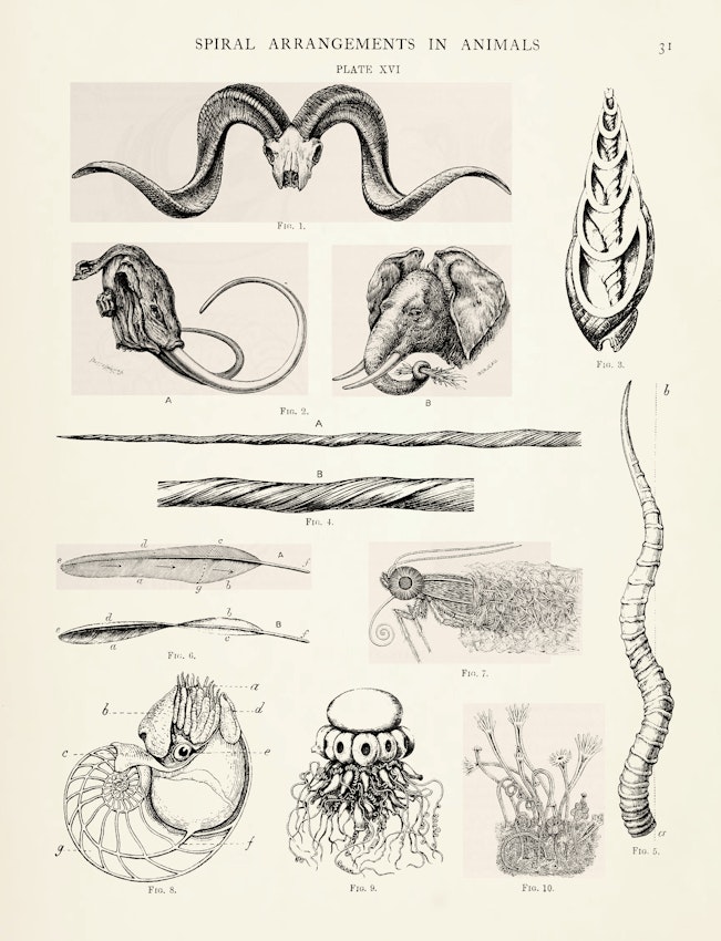 Scroll through the whole page to download all images before printing.
Plate XVI from the first volume of Pettigrew’s Design in Nature (1908), illustrating “spiral structures as seen in shells, horns, tusks, teeth, feathers, proboscides, tentacles, &c” — Source.
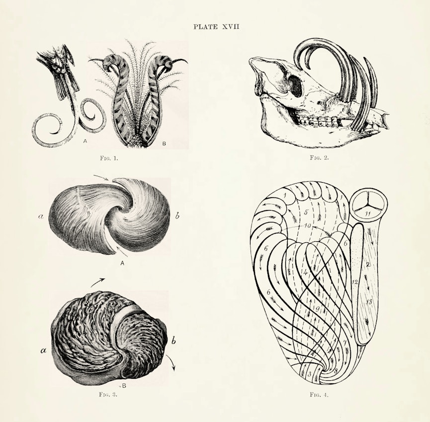 Scroll through the whole page to download all images before printing.
Plate XVII from the first volume of Pettigrew’s Design in Nature (1908), illustrating “spiral formations in feathers and teeth, in the muscular arrangements of the heart, and in the cast of the ventricular cavities of the heart” — Source.
At the center of this dizzying array lay the sacred secret that had so profoundly occupied Aristotle and Aquinas, Leonardo and Vesalius — the human heart. The heart’s sevenfold spiral structure was the mystery of mysteries, its form preserving perfectly the sense of both its muscular contractions and the interior circulatory patterns of its blood. Pettigrew had himself discovered this as a young medical student at the University of Edinburgh; so impressed had his professor been by Pettigrew’s dissections that he invited him to deliver the prestigious Croonian lecture at the Royal Society of London for 1860.
The University of Edinburgh was, in Pettigrew’s medical student days, at the zenith of its reputation: James Syme was dazzling the world with his bold pioneering surgery; James Young Simpson had — with dinner guests at his own 52 Queen Street table — proved the safety of chloroform as an obstetric anesthetic; with his methodical use of the microscope, John Hughes Bennett inaugurated a new era in the teaching of clinical medicine; Joseph Lister’s careful application of carbolic acid (phenol) to wounds, dressings, and instruments — though mocked initially by his medical colleagues — had revolutionized the practice of surgery.
When Pettigrew reminisced that the rivalry among these stellar physicians had been “a case of diamond cut diamond”, he recognized their fame by employing a most apt metaphor.7 Cutting — with a varied repertoire of scalpels, lancets, and scissors — was the surgeon’s special art. Pettigrew’s tutor in the art was Professor of Anatomy John Goodsir, who, with his large, powerful, finely shaped hands never failed to wield the scalpel “with a dexterity and grace truly remarkable”.8 At the end of the 1857–58 winter term, Professor Goodsir gave out as the subject for the senior anatomy gold medal: “The Arrangement of the Muscular Fibres in the Ventricles of the Vertebrate Heart”. This Gordian knot of anatomy had over the previous three centuries foiled the efforts of Vesalius, Albinus, Haller, and others to unravel it.9
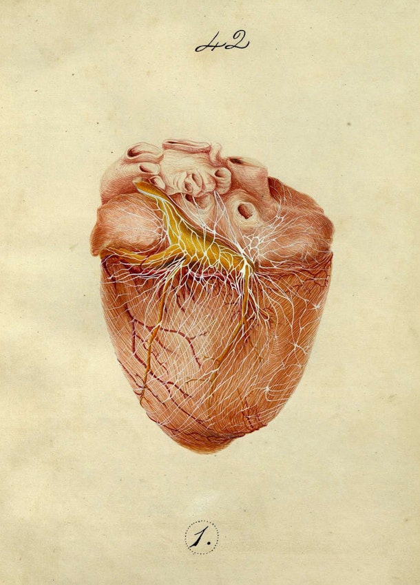 Scroll through the whole page to download all images before printing.
Dissection illustration from Pettigrew’s thesis on the “Arrangement of the cardiac nerves. . . in mammalia” — Source.
Back home, between sessions at his family’s country home in Lanarkshire, the twenty-four-year-old Pettigrew at once proceeded to dissect in large numbers every kind of heart within reach, making careful drawings and notes of each. Beginning with sheep, calf, ox, and horse, he found that he had to devise a new mode of dissection that would allow both sufficient hardness to preserve the anatomical structures, and ample softness so as to tease out their multitudinous tissue layers.
Having exhausted a battery of methylated spirits and other chemicals, he hit upon the expedient of stuffing and gently distending the ventricles of the heart with a truly Scottish material — dry oatmeal. Slowly boiling the hearts for four to five hours, he could get quit of all the external fat, blood vessels, nerves, lymphatics, and cellular tissue. A fortnight to three weeks hardening in a bath of methylated spirits followed, after which he was able to separate and peel off the muscular fibers of the ventricles as if they were layers of an onion. The layers were of two kinds: muscular fibers from the outside of the heart wound in a spiral direction from left to right, progressing downwards; the internal fibers ran in an opposite spiral direction from right to left, upwards.
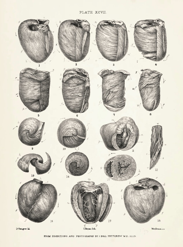 Scroll through the whole page to download all images before printing.
Plate XCVII from the second volume of Pettigrew’s Design in Nature (1908), illustrating the twisted and spiraling fibers of a heart — Source.
In fact, the internal and external layers of the muscular fibers of each and every one of the more than one hundred vertebrate hearts he had dissected formed two sets of opposite spirals which crossed each other, the crossings becoming more oblique towards the center. These inner and outer layers were further divided into a pair of left- and right-handed spiral sets. There was, especially in the left ventricle, a most perfect spiral symmetry, one that rivaled the Great Andromeda Nebula.
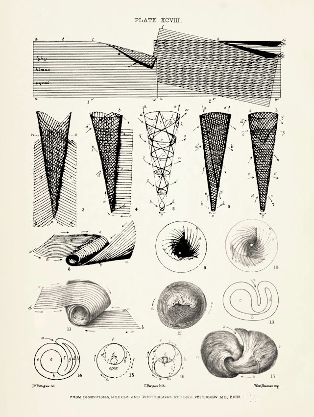
As gifted a model maker as he was a dissector, Pettigrew found this now-exposed double spiral heart to be an anatomical puzzle of the first order, for the external muscle fibers were seamlessly and spirally continuous with the internal muscle fibers at both the apex and base of the ventricles. One day Pettigrew came down to dinner a little earlier than usual, and, seeing a newspaper lying on the table, felt an impulse to roll it up obliquely from one corner — as grocers do in making conical paper bags. To Pettigrew’s surprise, the lines of print on the layers of newspaper ran in different directions according to a graduated order: the lines on the outer layers ran spirally from left to right downwards, becoming more oblique as the central layer was reached; the lines of print on the inner layer ran spirally from right to left upwards, becoming more vertical as they moved away from the center. The newspaper print on the two layers crossed at widening angles, forming an X, as the center was approached.
The print was seamless at both base and apex of the paper cone, resembling the arrangement of muscle fibers in the heart. There were, in effect, a series of complicated figure-of-eight loops, arranged in a marvelously mathematical pattern of great complexity and beauty. “Here”, he wrote, “was the whole thing in a nutshell. It was a case of the reading turning in or involuting at the apex and of the reading turning out or evoluting at the base”.10 Pettigrew’s newspaper model showed that the heart’s double helical structure — now known as the helical ventricular myocardial band (HVMB) — was essentially a triple-twisted Möbius strip.
 Scroll through the whole page to download all images before printing.
Plate XCVIII from the second volume of Pettigrew’s Design in Nature (1908) — Source.
Crying “EUREKA!”, Pettigrew ransacked the Lankarshire fish shops for the hearts of cod, salmon, sunfish, and turbot. He also lucked into securing the heart of a monster shark killed in the Firth of Forth. From the large hotels he collected several fine sea turtle hearts, as well as a land tortoise and an alligator. Raiding the poulterers too, he got the hearts of duck, goose, capercaillie, turkey, and one “splendid” swan’s heart.11
From fish to frog to turtle, the muscular fiber arrangement — though interesting — shed no light on the complicated arrangement in the ventricles of bird and mammal. (The pattern in the bird exactly matched that of the mammal except that, in the right ventricle of the bird, a muscular valve took the place of the fibrous tricuspid valve of mammals.) In the small hours of the morning, in his humble student lodgings, Pettigrew worked away now at dissections of sheep, calf, ox, horse, deer, pig, porpoise, seal, lion, giraffe, camel, and human — 112 dissections and associated drawings in all.
When the day for awarding the gold medal arrived, four hundred students crowded the large anatomical theater to hear the altogether unknown Pettigrew’s name pronounced. Professor Goodsir asked Pettigrew to call on him the next day, anxious to win the heart dissection preparations for the University’s Anatomical Museum. The 112 neat glass jars can still be found there today. He also invited young Pettigrew to report on his discoveries to the Royal Society of London; Pettigrew delivered his address, “On the Arrangement of the Muscular Fibres in the Ventricles of the Vertebrate Heart”, to the Royal Society the very same week that Origin of Species was published by John Murray of Albemarle Street, a short walk from the Royal Society lecture hall. That anyone might attribute such an ingeniously crafted organ as the mammalian heart to mere chance, Pettigrew believed, was sheer madness.
Nature’s variegated spiral structures, with the mammalian heart always for him the epitome, represented but one panel of the triptych that Pettigrew would go on to assemble over the next half century. Volume Two of Design in Nature is devoted solely to spiral movement in circulation (although the circulation section dealt with both plant and lower animal circulatory systems, three-quarters of this study focused on mammals and man); Volume Three to the spiral as locomotion’s characteristic form. In both arenas of animal physiology, Pettigrew found a spectacular resonance: movement at once precedes and follows structure, the direction of movement in living things being in every instance determined by the composition and configuration of kinetic spiral parts. This resonance seemed to reach right down to the atomic level. Unlike the closed system of the heart, the spiraling lines of atoms and molecules were arranged so that matter could be added in any amount, in unlimited directions. An open flow of energy and form was the basis for growth and progression in all creatures.
 Scroll through the whole page to download all images before printing.
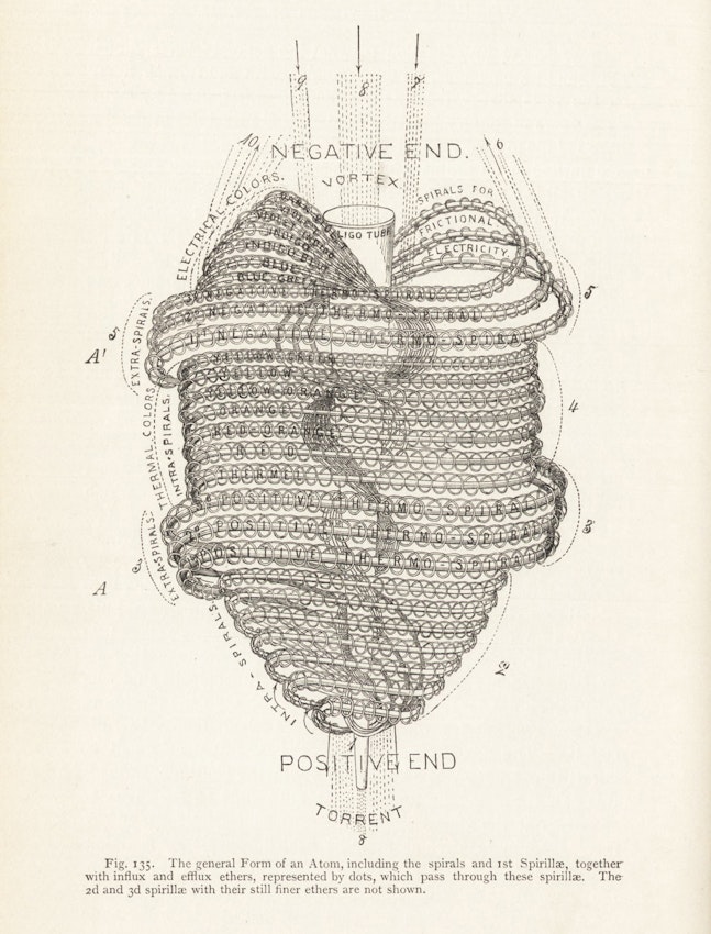
Illustration of an atom (looking remarkably like a heart), from Edwin D. Babbitt’s The Principle of Light and Color (1878). Note the labelled positive and negative spirals — Source.
Reflected in the vertebrate skeleton, this open attitude also made graceful locomotion possible. Pettigrew quoted his mentor John Goodsir: “The peculiar spiral attitudes into which the human body can be thrown are explained by the spiral curve of the vertebral articular surfaces, and the spiral arrangement of the muscles. No mammal can throw its trunk into those spiral curves which subserve the balance of the human frame and confer the peculiar grace and expression of its movements.”12 Only birds — especially his beloved swallows and swifts, which darted round the turrets of Swallowgate, the stone residence that Pettigrew had built at St. Andrews, and across the broad moor leading to the nearby sea cliffs — could rival the poetry of motion executed by the human body, their movements freed in the less resistant medium of air. The earthbound human body’s idiosyncratic spiraling structure liberated the hands to sculpt clay, tie rope, and grasp chalk, paintbrush, and scalpel in order to go inside the organs of Life and then represent them in color and line. Bony spirals hidden beneath spiral muscles flexed and extended to skip, leap, creep, crawl, wriggle, tumble, skate, march, flip, prance, moonwalk. The polka-ing, pirouette-ing, schottish-ing, waltzing, two-stepping human danced upon a near infinity of corporeal eddies.
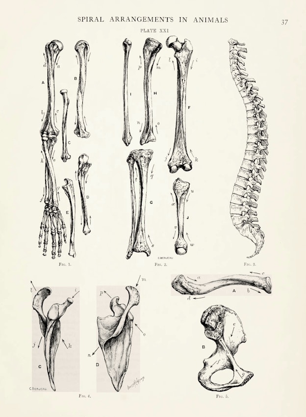
When Pettigrew took up the third strand of his argument from design, he began again with structures — the muscular and osseous systems, which he found intimately complimentary. Skeletal plates suggest that each part of our bony frame was but a partial realization of the sort of spiral geometry Pettigrew had discovered in the heart. The halfway twisting femur, humerus, tibia, fibula, ulna, and radius reached their fully spiral apotheosis in clavicle, pelvis, and scapula — each of which approached once again the geometry of the Möbius strip.
 Scroll through the whole page to download all images before printing.
Plate from the first volume of Pettigrew’s Design in Nature (1908), illustrating “the spiral formation of human bones as seen in the arm, leg, vertebral column, clavicle, scapula, and pelvis” — Source.
Published posthumously, just months before the centennial of Darwin’s birth, and the fiftieth anniversary of On the Origin of Species — celebrations which were fully exploited by Darwinians to advance a false picture of Darwin as a rabid opponent of teleology13 — Design in Nature dropped from sight as rapidly as Pettigrew’s ornithopter dropped from the sky over the St. Andrews moorland. Nature’s full-page review savaged the work’s teleological argument, wanly submitting that had Pettigrew lived to complete the editing of his opus, he would have “expunged or modified” its conclusions.14 Biostatistician Raymond Pearl was less generous, calling “the ponderous work” “probably the most extensive and serious single contribution to humorous literature which has appeared in recent years”, and declaring Pettigrew’s “spiral philosophy” — that “the Creator fashioned men and corkscrews on the same plan” — as “medieval as any cathedral”.15 Not a single work of contemporary biology or natural history — including D’Arcy Wentworth Thompson’s On Growth and Form (1917), which devoted considerable discussion to spiral forms — cited Design in Nature. A century on from its publication, however, Design in Nature holds up not only as an unsurpassed survey of biological form, but as a provocative modern inquiry into the causation of form. Pettigrew’s magnum opus is also a phenomenological masterpiece whose lively prose and gorgeous illustrations might serve to inspire a new generation of “spiralists”.
Kevin Dann’s books include Enchanted New York: A Journey Along Broadway Through Manhattan’s Magical Past (2020) and Expect Great Things: The Life and Search of Henry David Thoreau (2017). He leads extra-vagant bike tours in New York City. More at: www.drdann.com.


